Tricking taste buds but not the brain: Weekly consumption of artificial sweeteners changes the brain’s pleasure response to sweet treats

Originally published at Scientific American.

Do NOT EAT the chemicals. It is the #1 laboratory safety rule young scientists learn to never break and for good reason; it keeps lab citizens alive and unscathed. However, if it hadn’t been for the careless, rule-breaking habits of a few rowdy scientists ingesting their experiments, many artificial sweeteners may never have been discovered.

Perhaps the strangest anecdote for artificial sweetener discovery, among tales of inadvertent finger-licking and smoking, is that of graduate student Shashikant Phadnis who misheard instructions from his advisor to ‘test’ a compound and instead tasted it. Rather than keeling over, he identified the sweet taste of sucralose, the artificial sweetener commonly known today as Splenda.

Artificial sweeteners like Splenda, Sweet’N Low, and Equal provide a sweet taste without the calories.  Around World War II, in response to a sugar shortage and evolving cultural views of beauty, the target consumer group for noncaloric sweetener manufacturers shifted from primarily diabetics to anyone in the general public wishing to reduce sugar intake and lose weight. Foods containing artificial sweeteners changed their labels. Instead of cautioning ‘only for consumption by those who must restrict sugar intake’, they read for those who ‘desire to restrict’ sugar.

Today, the country is in the middle of a massive debate about the health implications of artificial sweeteners and whether they could be linked to obesity, cancer, and Alzheimer disease. It’s a good conversation to have because noncaloric sweeteners are consumed regularly in chewing gums, frozen dinners, yogurts, vitamins, baby food, and particularly in diet sodas.  As research delves deeper into these issues, scientists are gaining a greater understanding of how these sweet synthetic alternatives impact the brain. From engagement mechanisms of the brain’s central taste pathways, to uniquely altering the food reward-system response, we are learning that substituting one sweet taste for another by switching from sugar to artificial sweetener does not fool the brain. This brilliant organ knows the real deal even if your taste buds can’t detect the difference.

Sugar processing in the brain

The moment sugar touches your mouth a complex cascade of events is triggered involving taste, learning, memory, and reward systems in the brain.

The central taste pathway begins with your tongue, which has specialized cells that relay information about taste through cranial nerves to the brain. Taste information is then transmitted through several brain regions before arriving in the primary taste cortex, which is made up of the frontal operculum and the anterior insula. Neurons in the primary taste cortex send projections to areas associated with the brain’s primary reward-pathway located in the dopaminergic midbrain. Neurons within the midbrain then go on to innervate various brain centers that participate in the food reward response (i.e. amygdala, caudate nucleus, and orbitofrontal cortex) and release dopamine, a neurotransmitter commonly associated with reward and pleasure.

The body’s food-reward system plays a critical role in regulating eating behavior and controlling the number of calories you consume. Evolutionary survival mechanisms in the brain place emphasis on the value of high calorie foods and thus we find sugar satisfying so that we will continually seek it out.

The first bite of cupcake is always the best

Say you have a box of cupcakes. The initial bite is bliss. In that first taste, dopamine is released in the brain’s reward pathway and you get a jolt of pleasure. In addition to dopamine, the release of leptin, a hormone that regulates appetite and informs the brain when you are full, reduces activation of dopamine neurons in the midbrain, lowering the reward value of sugar. As a result, the second bite of the cupcake is less rewarding than the first and you begin to feel full with subsequent bites, hopefully stopping you from gorging yourself on the entire box of cupcakes.

What if instead you ate an artificially sweetened cupcake? Does indulging in artificially sweetened food and drink impact the central-taste and reward pathways in the brain? Functional magnetic resonance imaging (fMRI) studies have investigated this question and revealed some interesting findings.

Artificial sweeteners taste sweet but are not as rewarding to the brain as sugar

In a study conducted by Frank et al., 12 healthy women underwent brain scans and were asked to rate the pleasantness and sweetness of several different sugar (sucrose) and artificial sweetener (sucralose) drinks on a scale of 1 (‘did not like the taste’) to 9 (‘extremely enjoyable’).

Researchers found that both sugar and artificial sweetener activate the primary taste pathway in the brain by activating the frontal operculum and the insula, but only real sugar was able to elicit a significant response from several brain regions of the taste-reward system including the midbrain and caudate nucleus. This suggests that the brain’s reward pathway is conditioned to prefer a sugar, or caloric-based, stimulus.

But what happens if you routinely drink diet soda? If sweet taste is no longer a reliable measure of caloric intake because you regularly consume artificial sweeteners, does the brain’s reward response to sweet taste change? Potentially yes, and here’s why:

At the San Diego State University, researchers recruited 24 individuals for an fMRI study to look at brain activation of habitual diet soda drinkers and non-diet soda drinkers. Study participants were grouped as diet soda drinkers if they drank at least one diet beverage a week. On average, diet soda drinkers in the study consumed 8 diet beverages a week.

During the brain scan, subjects were provided with random intermittent sips of sugar (sucrose) water and artificially sweetened (saccharin) water. After each trial taste, they were asked to rate drink pleasantness and given distilled water to cleanse their palate before the next trial.

Green and Murphy found that chronic diet soda drinkers had greater overall activation in several reward processing brain regions to both real sugar and artificial sweetener, compared to the non-diet soda group. Additionally, within diet soda drinkers, the brain’s response to sugar vs. artificial sweetener was nearly identical in the orbitofrontal cortex, dopaminergic midbrain, and amygdala, suggesting regular consumption of diet soda may render particular components of the brain’s reward system incapable of distinguishing between real sugar and artificial sweetener!

Furthermore, while certain components of the reward pathway were numb to sweet taste type, researchers found that the more diet soda an individual consumed, the lower their activation was in the caudate nucleus. Thus, people that drank the most diet soda had the least activity in the caudate head region.

Taste and reward signaling in the brain is immensely complex. Research is only beginning to understand how altered brain activity with prolonged use of artificial sweeteners may impact our health long-term. While previous studies have shown an association between obesity and decreased caudate head activation during food-reward tasks, a link between artificial sweeteners altering brain activity in the caudate head and obesity has not yet been established. Future fMRI studies as well as looking at how appetite hormones, like leptin, alter the brain’s reward pathway after regular use of artificial sweeteners could further piece together this incomplete picture.

Even if you aren’t married to the clean eating fad, the take home message is that real sugar or not, moderation is key for a healthy brain-reward response. Or as Cookie Monster with his new health-motivated outlook might put it: cupcakes are a sometimes food.

Green E. & Murphy C. (2012). Altered processing of sweet taste in the brain of diet soda drinkers, Physiology & Behavior, 107 (4) 560-567. DOI:

Frank G.K.W., Oberndorfer T.A., Simmons A.N., Paulus M.P., Fudge J.L., Yang T.T. & Kaye W.H. (2008). Sucrose activates human taste pathways differently from artificial sweetener, NeuroImage, 39 (4) 1559-1569. DOI:

Image credit: Roadsidepictures (via Flickr)

Being Hangry: The neuroscience behind hunger and a sour mood

Where are our meals? The service at this restaurant is awful. We’ve been waiting for an hour and have yet to even see a glimpse of our appetizers! Those people ordered after us and they just got their food! I’m starving! Don’t tell me to relax; I’m starting to get hangry!

Eek! Sound like an all too familiar scenario? “Hanger”, the portmanteau or mash-up of the words hungry and anger describing a state of rage caused by lack of food, may actually be linked to levels of the neurotransmitter serotonin in the brain.

Be kind, consume some glucose

Photo credit: Carlos Santa Maria

Photo credit: Carlos Santa Maria

What happens to our mood when our body is running low on glucose a.k.a. sugar? Researchers at the University of Kentucky were interested in the link between low glucose levels and aggressive behavior, so they designed a devious study to investigate the sugar-mood association.

In the study, 62 college students were asked to drink lemonade containing either sugar or a sugar substitute. After drinking their randomized beverage, the students participated in a “game” where they were told that they were competing against an opponent to see who could press a button the fastest.

As it turns out, the whole thing was rigged. There was no opponent just a computer. The students were set-up from the beginning to lose about 50% of the time. The loser of each round would receive a blast of white noise in their headphones. Ouch! Additionally, before each new round, the student selected the level and duration of noise their “opponent” would receive following a loss on that round.

As students began receiving white-noise blasts after “losses”, they retaliated, as any frustrated person might do, and tried to return the favor to their opponent by matching the white-noise assault. Interestingly, researchers found that when students were provided with a sugar-substitute lemonade (no glucose) they were more aggressive, providing louder and longer noise blasts, than if they drank the lemonade with sugar. Feeling agitated? Have a glass with glucose and chill out!

How low blood sugar impacts the brain

Your brain needs fuel in order to function properly. Most often this fuel comes in the form of glucose. When you go several hours without eating, your blood sugar drops. Once it falls below a certain point, glucose-sensing neurons in your ventromedial hypothalamus, a brain region involved in feeding, are notified and activated resulting in level fluctuations of several different hormones. Ghrelin, a hormone that increases expression when blood sugar gets low and stimulates appetite through actions of the hypothalamus, has been shown to block the release of the neurotransmitter serotonin. The serotonin system is incredibly complex and contributes to a number of different central nervous system functions. One of the many hats this neurotransmitter wears is modulation of emotional state, including aggression.

Is your mood more difficult to control when serotonin is depleted?

Angry and neutral faces during the task. Brain regions impacted following serotonin depletion: vACC - ventral anterior cingulate cortex; VLPFC - ventrolateral prefronal cortex. (Passamonti et al. 2011)

Angry and neutral faces during the task. Brain regions impacted following serotonin depletion: vACC – ventral anterior cingulate cortex; VLPFC – ventrolateral prefronal cortex. (Passamonti et al. 2011)

Potentially yes, and here’s why. In an functional magnetic resonance imaging (fMRI) study, Passamonti et al. looked at how neuronal networks involved in processing aggression were altered in subjects with low serotonin levels. Nineteen healthy participants underwent brain scans on two separate days: once after consuming a tryptophan-depleting drink and again after drinking a placebo beverage containing tryptophan. Tryptophan is an essential amino acid, found in turkey among other protein sources, that is a building block for serotonin formation.  Your body does not make tryptophan on it’s own and you must get it through your diet. Don’t worry though, there’s plenty of it around! By reducing tryptophan levels, researchers were able to evaluate the effects of low serotonin levels on brain connectivity in individuals viewing angry faces.

After serotonin depletion, participants were scanned to assess brain responses to images of angry, sad, and neutral faces that were presented to them.  Participants were also asked to complete a personality questionnaire to evaluate their individual propensity for aggression.

What did they find? By reducing serotonin through tryptophan depletion, the connectivity between the amygdala and two prefrontal cortex regions, the ventral anterior cingulate cortex and the ventrolateral prefrontal cortex, was altered when processing angry faces but not sad or neutral faces.

Additionally, when looking at individuals that were more prone to aggression based on their personality questionnaires, their brain scans revealed weaker connections between the amygdala and the prefrontal cortex.  Meaning if you have a predisposition to aggression, low serotonin levels circulating in your brain may lead to altered communications between brain regions that wrangle aggressive behavior.

Angry at a restaurant? Stuck in traffic? Late for dinner and feeling a Dr. Jekyll and Mr. Hyde scenario about to unfold? It may be due to serotonin messing with your brain. Grab your emergency turkey sandwich and relax. Life is going to be okay.

DeWall C.N., Deckman T., Gailliot M.T. & Bushman B.J. (2011). Sweetened blood cools hot tempers: physiological self-control and aggression, Aggressive Behavior, 37 (1) 73-80. DOI:

Passamonti L., Crockett M.J., Apergis-Schoute A.M., Clark L., Rowe J.B., Calder A.J. & Robbins T.W. (2011). Effects of acute tryptophan depletion on prefrontal-amygdala connectivity while viewing facial signals of aggression., Biological psychiatry, PMID:

Tingling palms and knocking knees: Why do we fear heights?

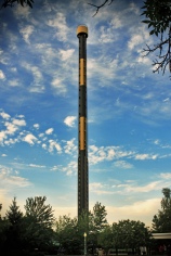
Kennywood – Pittsburgh’s premier amusement park – has filled my childhood with magical memories. Riding the stunning carousel horses around-and-around to the accompaniment of big band music. The scent of Potato Patch fries and funnel cake wafting through air. But what is the one memory I’ll never forget? The 251-foot Pitfall scaring the living daylights out of me.

The Pitfall at Kennywood Park

The Pitfall at Kennywood Park. Photo credit: Scott Jones

The Pitfall, now a retired ride, would leisurely take you 251 feet up into the sky and pause at the top so you could take in the view. With nothing more than a subtle *click* the brakes release and in a brief terrifying moment riders scream as they plummet towards earth.

One year during my 7th grade Kennywood school picnic, in a heroic effort I convinced some nervous friends that going on the Pitfall was the best idea ever. A few were crying actual tears in protest but ultimately we decided to ride. You’re only in 7th grade once, right?

We got in line and when our turn arrived a park employee locked us down in our seats, legs dangling below us. I had built the Pitfall up so much in my mind and I was confident about our decision to ride until we made it halfway up the ascent.

Halfway up. That’s where the whole idea turned sour. Turns out I have acrophobia – I’m deathly afraid of heights!

 Just looking at this picture makes my hands start to tingle!

The neurobiology of fear

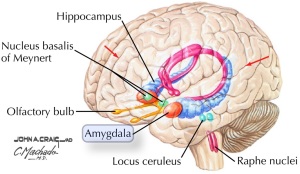
Where does fear originate in our brain? Scientists believe that the brain region known as the amygdala plays an important role in triggering fear. Greek for ‘almond’, describing it’s shape, the amygdala sits in the medial temporal lobe of the brain.

The amygdala highlighted in orange.

The amygdala highlighted in orange.

A fear stimulus, such as heights, activates a cascade of events in the brain. The sensory cortex acknowledges something as frightening and signals the amygdala. In turn, the amygdala notifies the hypothalamus and the brainstem and you feel fear.

In humans, a very rare hereditary illness, Urbach-Wiethe disease, can cause bilateral symmetrical loss of all the cells in the amygdala. Without the amygdala the fear signaling cascade is broken and individuals are unable to experience fear. One famous patient, a 44-year-old woman known only as SM, exemplifies this fearlessness.

One night while walking home past a park, SM was grabbed by a man who put a knife to her throat, and exclaimed, “I’m going to cut you!”. Without feeling afraid she replied, “If you’re going to kill me, you’re gonna have to go through my God’s angels first.” Ballsy and effective. He responded by releasing her and she calmly walked back home. Extraordinarily, the following night she walked past the very same park again!

SMBrainScanMRI brain scan of SM compared to a healthy control subject.
The regions circled in red represent the amygdala and in SM is void of tissue.

Like others with Urbach-Wiethe disease, SM has characteristic bilateral amygdala lesions. SM’s IQ, memory, and language are normal. Although she experiences a wide range of other emotions, in her adult life there has never been an instance in which she has felt fear.  She appears to understand the concept of fear, having personally felt it once as a child, when she was cornered by a friend’s Doberman Pinscher. This incidence was presumably prior to loss of her amygdala. As an adult SM is able to recognize fear from body language and prosody of an individual’s voice, but interestingly she is unable to discern fear in static facial expressions.

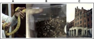
In a study by Feinstein et al., SM was asked to participate in a number of frightening tasks. She was shown terrifying horror films, asked to hold large snakes and spiders, and taken to a haunted house. She never once showed signs of fear and when prompted described her experiences as feeling overwhelmingly “curious”.

SMFear(A) SM holding a snake,
(B) A spider SM tried to touch,
(C) Waverly Hills Sanatorium Haunted House SM toured

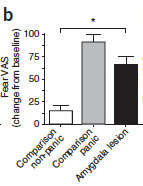
Self-described levels of fear following carbon dioxide inhalation.

Self-described levels of fear following carbon dioxide inhalation.

In a new study Feinstein and colleagues tried a different approach to induce fear and panic in SM and two other subjects with Urbach-Wiethe disease by exposing them to carbondioxide (CO2). Breathing in CO2 creates the sensation of suffocation and upon inhalation the subjects described feelings of being “overwhelmed by the panic and fear of dying”. It worked! SM felt fear for the first time in her adult life. But how?

The brainstem controls basic bodily functions such as breathing and heart rate. SM’s sensation of fear suggests that ultimately the brainstem, the endpoint for the fear cascade, holds the key to the conscious experience of fear. Specific threats, such as CO2, may bypass the circuit and impact the brainstem directly, eliciting fear without receiving a signal that has been processed through higher brain regions and the amygdala.

While scientists have not yet performed a field experiment with SM riding a Kennywood-escque Pitfall, Daniel Tranel, a professor of neurology and psychology at the University of Iowa, has been studying SM for years and tells NPR that SM reports being unafraid of heights. So unless she is sucking in CO2 on the ride, a simple roller coaster used as a fear stimulus that would be processed through the amygdala rather than directly impacting the brainstem, is unlikely to faze her.

Do we have an innate fear of heights?

Barring that you have an intact amygdala, are we programmed to fear heights from birth? Several studies have addressed this issue using a visual cliff.

Visual Cliff

A visual cliff is a trick-of-the-eye testing apparatus where an opaque patterned surface is connected to a transparent glass surface. Below the transparent side is a lower level that has the same pattern as the opaque surface. The visual cliff creates the illusion that you could fall over the edge.

Researchers noticed that if an infant was able to crawl and move on their own accord then they were also more wary of the “cliff”. However, if the child was “prelocomotor”, or younger than crawling age, they did not fear the edge. This is likely because as a baby learns to move around they also become aware of distances, depth perception, and begin coordinating their visual system with movement through their environment.

This experiment has been reproduced in a number of animal species including kittens that also utilize visual cues in movement. The visual cliff did not deter animals, such as rats, which predominately rely on tactile cues by whisking surfaces with their whiskers rather than vision to navigate their environment.

Thus it would seem as though fear of heights is a learned response to experiences, such as falling or near falling incidences, rather than something we are born with.

So what happened with the Pitfall after I realized that being more than 15 feet up in the air was too high for me? Well I survived. I actually rode the ride several more times since, rationalizing that the ride was engineered well and I was probably statistically safe.  If you ever get the chance to ride something like the Pitfall, try placing a dime on your knee to watch it levitate in front of you as you drop.  It might just keep your mind off of the heights.

Feinstein J.S., Adolphs R., Damasio A. & Tranel D. (2010). The human amygdala and the induction and experience of fear., Current biology : CB, PMID:

Feinstein J.S., Buzza C., Hurlemann R., Follmer R.L., Dahdaleh N.S., Coryell W.H., Welsh M.J., Tranel D. & Wemmie J.A. (2013). Fear and panic in humans with bilateral amygdala damage., Nature neuroscience, PMID:

Campos J.J., Bertenthal B.I. & Kermoian R. (1992). EARLY EXPERIENCE AND EMOTIONAL DEVELOPMENT: The Emergence of Wariness of Heights, Psychological Science, 3 (1) 61-64. DOI: