A secret to unlocking happiness? Practice gratitude.

Friends & family. Puppies. Pittsburghese. Sunday mornings. Rainy summer evenings. Hugs. Books. Driving with the windows down. The Beatles. Trail runs. Pumpkin patches. The brain.

Lots of things make me happy – but few things feel as wonderful as showing appreciation for the kindness, love, or courtesy extended by another person.

My Buddha Doodles Gratitude Journal by Molly Hahn (Mollycules)! I love the art in this journal and there is plenty of space provided to write down positive experiences from each day.

Gratitude, originating from the Latin word gratus meaning ‘thankful’, is a powerful moral sentiment that psychologists have shown significantly influences overall happiness.  In the simple act of being thankful, many mental, social, and physical benefits follow in tow.

Earlier this year I came across the SoulPancake video titled “The Science of Happiness – An Experiment in Gratitude”. The video explored a result from a 2005 study that investigated the effectiveness of several positive psychology interventions on prolonging happiness over several months. These interventions included exercises in gratitude and building positive self-awareness.

Part of the original 2005 study directed participants to complete a questionnaire that established their general level of happiness. Following the survey, they were instructed to write and deliver a letter of gratitude to someone who had been especially kind toward them but had not been properly thanked.

One week after the letter, participants reported being happier and less depressed. This particular exercise in reflection and expression of gratitude created the largest happiness boost out of all the activities assigned in the study.  In other words, telling someone how much you appreciate them produces the most happiness bang for your buck.

In addition, these positive vibes were maintained through the one-month study follow-up, but did not last until the 3-month follow-up – so practice gratitude often!

A separate group of study participants were given the task to journal about three good things that happened each day. This group reported being happier than before the study and they stayed happier at the 3- and 6-month study follow-ups. Additionally, the happiest people journaled about their good experiences frequently.

Hence, my 2014 New Years Resolution is keeping a gratitude journal. Bring on the gratitude glow!

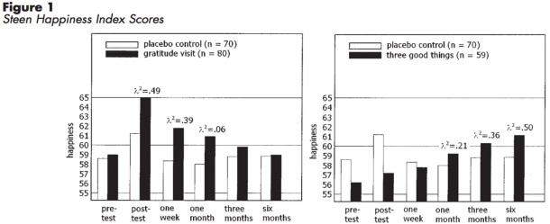
Gratitude letter writing (left) & journaling 3 good daily things (right)
effect on happiness over 6 months. Seligman et al. 2005

It may not be shocking, but grateful people take better care of themselves and see mental and physical benefits on top of enhanced happiness. Other studies have revealed that the practice of gratitude is associated with better sleep, decreased anxiety, and improved exercise habits.

Although with too little sleep, the positive effects of gratitude no longer benefit anxiety. So be grateful and get those nighttime Z’s!

Expressing gratitude has the ability to alter happiness and impact our physical well-being, but what brain centers respond to moral sentiments of gratitude?

In 2009, the NIH conducted an fMRI study assessing cerebral blood flow – thought to be a measure of neural activity in the brain – while subjects were shown positive word pairings to trigger feelings of gratitude. The more gratitude a participant  experienced, the more active their hypothalamus became. Because the hypothalamus is an important brain region for body temperature, sleep, metabolism, hunger, and the body’s stress response, it’s logical that expressing gratitude can lead to improvements in sleep, depression, and anxiety.

Gratitude. It’s the reason for the season. But in 2014 let’s celebrate and express gratitude not only during the holidays but frequently throughout the year. I’d like to start this year by saying how thankful I am to you,  The Synaptic Scoop readers. You make writing this blog a joy. Thank you and have a very Happy New Year filled with lots of gratitude!

Seligman M.E.P., Steen T.A., Park N. & Peterson C. (2005). Positive Psychology Progress: Empirical Validation of Interventions., American Psychologist, 60 (5) 410-421. DOI:

Zahn R., Moll J., Paiva M., Garrido G., Krueger F., Huey E.D. & Grafman J. (2009). The Neural Basis of Human Social Values: Evidence from Functional MRI, Cerebral Cortex, 19 (2) 276-283. DOI:

New Year, New Scoop!

Hello Synaptic Scoopers! It’s almost time to ring in the new year! I have a number of exciting things in store for 2014 including: guest posts, book reviews, a new blog look, collaborations, and of course more neuroscience scoop!

Until then, if the mood strikes you, check out these posts I’ve recently written over at BeingHuman.org:

The New Science of Cuffing Season

Gene expression modifies monogamy


Too Old for Santa Claus?

How age affects imagination


concert

Higher Status, Better Health

Social status alters gene expression


Have a very Happy New Year!

The neurobiology of “cuffing season” (How the brain influences monogamy)

Prairie Voles in LoveIt’s that time of year again – cuffing season! What’s cuffing season you ask? Well, according to UbranDictionary.com, cuffing season refers to the fall and winter months when normally promiscuous singles begin to look for serious relationships (becoming “cuffed” or tied down) due in part to colder weather and staying indoors. This seasonal phenomenon of pairing-up has inspired songs by John Mayer and Mumford and Sons, but why does it happen? Neurobiologists at Florida State University have identified some of the factors that may be responsible for “cuffing season” by studying prairie voles in love.

Prairie voles are a great animal model for studying the neurobiological underpinnings of social attachment because, aside from being adorable, they have a tendency to form enduring monogamous relationships.

In previous studies, researchers found that prairie voles preference for forming lasting pair-bonds was a result of a neurotransmitter love potion cocktail in the brain involving: oxytocin, vasopressin, and dopamine. Additionally, vole partner preference in female prairie voles was influenced by gene expression of oxytocin (OTR)  receptors in the nucleus accumbens, a brain region involved in reward and pleasure.

Knowing that gene expression caused changes in paring behavior, Wang and colleagues set out to determine if epigenetic modifications may also be contributing to the why behind “why do voles fall in love?”.

Histones wrapped in DNA

Histones wrapped by DNA
Illustration credit: Thom Graves

Epigenetics is a relatively new field of study looking at changes in gene expression caused by modifications in the availability of the DNA sequence to be read and transcribed. One such epigenetic modification known as histone deacetylation (HDAC) involves histones, which are the globs of protein used by cells to neatly package incredibly long segments of DNA by coiling it around and around. When DNA is tightly wound around histones, the histones take on a unique role in gene regulation by physically varying the accessibility of certain segments of DNA for expression. Think about wrapping yarn around a basketball. If the yarn is tight you can’t fit anything between the string and the ball. In the case of DNA, if it is tightly wound around the histone the cell machinery that reads the nucleic acid sequence cannot do its job. During HDAC, the removal of an acetyl group bound to the histone causes the DNA to condense and coil up further, reducing levels of gene transcription.

Wang et al. used HDAC inhibitors that allow histones to loosen their grip on DNA, enhancing gene expression, to see if adult female prairie voles changed their preference for their partner or if they would decide to spend time with a total stranger.

Did these HDAC inhibitors make a prairie vole love connection?

Sure did. When prairie voles were given HDAC inhibitors there was a significant increase in the amount of time they spent side-by-side touching their partner.

But what was the neurobiological action of the HDAC inhibitors?

To assess what might be happening down at the molecular level, researchers first looked at whether there were changes in OTR and and vasopressin receptor (V1aR) expression following HDAC inhibition. When HDAC inhibitors were administered, OTR mRNA and protein levels in the nucleus accumbens were significantly elevated. V1aR protein levels, although not mRNA levels, were also increased in this brain region.

Researchers then took it a step further to see if the genes for each receptor were more actively transcribed leading to the creation of more protein following HDAC inhibition and decided to look at histone acetylation in the OTR and V1aR gene promoter region, a region of DNA that initiates transcription of a gene. Following administration of HDAC inhibitor, there was a significant increase in histone acetylation in both receptors’ promoter regions suggesting that epigenetic modification was impacting behavior specifically at the promoter site.

To ensure that this finding was specific to OTRs and V1aRs in the nucleus accumbens these receptors were pharmacologically blocked. When either of these receptors no longer functioned, the HDAC inhibitors effect was diminished and prairie voles stopped caring whether or not they spent time with the love of their life or a complete stranger.

Finally, researchers found that mating triggered the same OTR and V1aR mechanism of action on gene promoters as administering HDAC inhibitors in the laboratory, reinforcing idea that in nature epigenetics do play a part in the neurobiology of monogamous relationships.

Now I definitely don’t recommend trying to go inject that cutie with a booty you’ve had your eye on with HDAC inhibitors in attempt to woo them as your cuffing season anxieties set in. That would definitely land you with jail time and perhaps give cuffing season a completely new meaning. If you really want to meet someone as the leaves start to change and the nights get cooler, remember a much better way to go is with confidence and a smile.

Wang H., Duclot F., Liu Y., Wang Z. & Kabbaj M. (2013). Histone deacetylase inhibitors facilitate partner preference formation in female prairie voles, Nature Neuroscience, 16 (7) 919-924. DOI:

Image credit: The McGraw Lab at NCSU

Flavor is all in your head: An amalgamation of senses created in the brain, not a property of food

Most people have probably never sat around and meditated deeply on the origin of potato chip flavor. But if you really begin to think about it, most of what we perceive as flavor is not mouth-derived but actually due to your sense of smell.

The Jelly Bean Experiment is a clever DIY test to help illustrate this point. All you need is a friend and a bag of jelly beans.

Got your supplies? Good.

Now close your eyes and have your friend select a bean for you. See if you can detect the flavor. Do this with a few different kinds of beans. Once you’ve established that you have a sense of taste (it’s not a test to see if you can become the next connoisseur of jelly beans, just that you can tell two flavors apart), with your eyes still closed, pinch your nose and have your friend hand you another bean. Can you tell what flavor it is? Probably not. You can most likely detect sweetness but your ability to distinguish the flavor of the bean is gone.

Distinguishing flavors is a common issue for people with anosmia, or the inability to smell. While great for the smellier parts of life, like gym socks or baby diapers, anosmia makes detecting the subtle nuances of sage, basil, and oregano in Grandma’s homemade pasta sauce a hopeless endeavor. Without smell, it’s not going to happen.

Reduced sniffer capability is also partially responsible for airline food being so troublingly tasteless. Fellow science writer Jordan Gaines Lewis wrote a great piece on many of the reasons why airline food is unpleasant with some fault laying with your olfactory (smell) system – Check it out HERE!

Retronasal smell through exhaling air. Shepherd, Nature 2006.

Retronasal smell through exhaling air. Shepherd, Nature 2006.

Smell all the smells and watch as each odor molecule uniquely engages your olfactory system

Let’s say that you’re in the laundry room trying to decide if a sock needs washed. When you breathe in the essence of the sock through your nose (hopefully its essence is spring rain, not dirty shoe), you are using orthonasal stimulation. However, there is a second smell pathway that allows you to detect scents from inside your body, referred to as retronasal stimulation. This sounds gross but is the process that actually occurs when you are chewing food and exhale.

In either direction, orthonasal or retronsal, odor molecules come in contact specialized tissue in the nasal cavity called the olfactory epithelium. The neurons within the olfactory epithelium are activated and send signals to the olfactory bulb.

Here’s where things get kind of crazy. Each smell, or odor molecule, has a unique chemical structure that results in a unique neuronal activity pattern in the olfactory bulb. When imaging how different chemicals affected the olfactory bulb in a mouse, researchers found that by just adding one carbon atom to a molecule, the ‘odor image’ on the bulb changed, meaning each smell showed a distinctive pattern of activation. Because the olfactory system can distinguish between single-carbon differences it is one of the finest molecular discriminators in the nervous system!

Heat map of olfactory bulb activation in response to different odor molecules.
Red indicates ares of highest activation. Notice how each molecule (shown below the bulb)
changes the activation response. They only differ by number of carbon atoms!

Once the olfactory bulb is activated, the neurons in the bulb send signals up through several brain regions continuing to the olfactory cortex, located in the orbitofrontal cortex. The orbitofrontal cortex is a common meeting ground in the brain where several different brain sensory modalities converge. It is through these multi-sensory inputs that the experience of taste is created.

Flavor processing is immensely complex because it engages almost all of your senses at once

So returning to the wonderment that is the potato chip eating experience – how is flavor being created in the brain? By just opening the bag of chips you’re visual system kicks it into high gear, mobilizing your gut as you begin to anticipate the food and salivate. Once the chip is in your mouth, taste, auditory, and motor systems are triggered as you begin to audibly crunch up the snack. Retronasal smell impacts your olfactory system as you chew and breathe. The brain is integrating all of these sensory factors and assembling the sour cream and onion flavor you have come to know, love, and savor.

While smell is likely one of the dominant sensory modalities in flavor creation, the visual system has also been shown to play a critical role. One study asked both novice and experienced wine testers to classify red and white wines by taste. The trick was that researchers colored some of the white wine with a red dye. They found that people classified wines by color rather than by taste showing, at least in this scenario, that the information from the visual system was able to override olfactory information in determination of taste.

To learn more about the “human brain flavor system” check out the book Neurogastronomy: How the Brain Creates Flavor and Why It Matters by Gordon M. Shepherd, Yale neuroscientist and head honcho in this field.

Shepherd G.M. (2006). Smell images and the flavour system in the human brain, Nature, 444 (7117) 316-321. DOI:

Xu F. (2003). Odor maps of aldehydes and esters revealed by functional MRI in the glomerular layer of the mouse olfactory bulb, Proceedings of the National Academy of Sciences, 100 (19) 11029-11034. DOI:

Morrot G., Brochet F. & Dubourdieu D. (2001). The Color of Odors, Brain and Language, 79 (2) 309-320. DOI:

Image credit: Keith Williamson (via Flickr

Tricking taste buds but not the brain: Weekly consumption of artificial sweeteners changes the brain’s pleasure response to sweet treats

Originally published at Scientific American.

Do NOT EAT the chemicals. It is the #1 laboratory safety rule young scientists learn to never break and for good reason; it keeps lab citizens alive and unscathed. However, if it hadn’t been for the careless, rule-breaking habits of a few rowdy scientists ingesting their experiments, many artificial sweeteners may never have been discovered.

Perhaps the strangest anecdote for artificial sweetener discovery, among tales of inadvertent finger-licking and smoking, is that of graduate student Shashikant Phadnis who misheard instructions from his advisor to ‘test’ a compound and instead tasted it. Rather than keeling over, he identified the sweet taste of sucralose, the artificial sweetener commonly known today as Splenda.

Artificial sweeteners like Splenda, Sweet’N Low, and Equal provide a sweet taste without the calories.  Around World War II, in response to a sugar shortage and evolving cultural views of beauty, the target consumer group for noncaloric sweetener manufacturers shifted from primarily diabetics to anyone in the general public wishing to reduce sugar intake and lose weight. Foods containing artificial sweeteners changed their labels. Instead of cautioning ‘only for consumption by those who must restrict sugar intake’, they read for those who ‘desire to restrict’ sugar.

Today, the country is in the middle of a massive debate about the health implications of artificial sweeteners and whether they could be linked to obesity, cancer, and Alzheimer disease. It’s a good conversation to have because noncaloric sweeteners are consumed regularly in chewing gums, frozen dinners, yogurts, vitamins, baby food, and particularly in diet sodas.  As research delves deeper into these issues, scientists are gaining a greater understanding of how these sweet synthetic alternatives impact the brain. From engagement mechanisms of the brain’s central taste pathways, to uniquely altering the food reward-system response, we are learning that substituting one sweet taste for another by switching from sugar to artificial sweetener does not fool the brain. This brilliant organ knows the real deal even if your taste buds can’t detect the difference.

Sugar processing in the brain

The moment sugar touches your mouth a complex cascade of events is triggered involving taste, learning, memory, and reward systems in the brain.

The central taste pathway begins with your tongue, which has specialized cells that relay information about taste through cranial nerves to the brain. Taste information is then transmitted through several brain regions before arriving in the primary taste cortex, which is made up of the frontal operculum and the anterior insula. Neurons in the primary taste cortex send projections to areas associated with the brain’s primary reward-pathway located in the dopaminergic midbrain. Neurons within the midbrain then go on to innervate various brain centers that participate in the food reward response (i.e. amygdala, caudate nucleus, and orbitofrontal cortex) and release dopamine, a neurotransmitter commonly associated with reward and pleasure.

The body’s food-reward system plays a critical role in regulating eating behavior and controlling the number of calories you consume. Evolutionary survival mechanisms in the brain place emphasis on the value of high calorie foods and thus we find sugar satisfying so that we will continually seek it out.

The first bite of cupcake is always the best

Say you have a box of cupcakes. The initial bite is bliss. In that first taste, dopamine is released in the brain’s reward pathway and you get a jolt of pleasure. In addition to dopamine, the release of leptin, a hormone that regulates appetite and informs the brain when you are full, reduces activation of dopamine neurons in the midbrain, lowering the reward value of sugar. As a result, the second bite of the cupcake is less rewarding than the first and you begin to feel full with subsequent bites, hopefully stopping you from gorging yourself on the entire box of cupcakes.

What if instead you ate an artificially sweetened cupcake? Does indulging in artificially sweetened food and drink impact the central-taste and reward pathways in the brain? Functional magnetic resonance imaging (fMRI) studies have investigated this question and revealed some interesting findings.

Artificial sweeteners taste sweet but are not as rewarding to the brain as sugar

In a study conducted by Frank et al., 12 healthy women underwent brain scans and were asked to rate the pleasantness and sweetness of several different sugar (sucrose) and artificial sweetener (sucralose) drinks on a scale of 1 (‘did not like the taste’) to 9 (‘extremely enjoyable’).

Researchers found that both sugar and artificial sweetener activate the primary taste pathway in the brain by activating the frontal operculum and the insula, but only real sugar was able to elicit a significant response from several brain regions of the taste-reward system including the midbrain and caudate nucleus. This suggests that the brain’s reward pathway is conditioned to prefer a sugar, or caloric-based, stimulus.

But what happens if you routinely drink diet soda? If sweet taste is no longer a reliable measure of caloric intake because you regularly consume artificial sweeteners, does the brain’s reward response to sweet taste change? Potentially yes, and here’s why:

At the San Diego State University, researchers recruited 24 individuals for an fMRI study to look at brain activation of habitual diet soda drinkers and non-diet soda drinkers. Study participants were grouped as diet soda drinkers if they drank at least one diet beverage a week. On average, diet soda drinkers in the study consumed 8 diet beverages a week.

During the brain scan, subjects were provided with random intermittent sips of sugar (sucrose) water and artificially sweetened (saccharin) water. After each trial taste, they were asked to rate drink pleasantness and given distilled water to cleanse their palate before the next trial.

Green and Murphy found that chronic diet soda drinkers had greater overall activation in several reward processing brain regions to both real sugar and artificial sweetener, compared to the non-diet soda group. Additionally, within diet soda drinkers, the brain’s response to sugar vs. artificial sweetener was nearly identical in the orbitofrontal cortex, dopaminergic midbrain, and amygdala, suggesting regular consumption of diet soda may render particular components of the brain’s reward system incapable of distinguishing between real sugar and artificial sweetener!

Furthermore, while certain components of the reward pathway were numb to sweet taste type, researchers found that the more diet soda an individual consumed, the lower their activation was in the caudate nucleus. Thus, people that drank the most diet soda had the least activity in the caudate head region.

Taste and reward signaling in the brain is immensely complex. Research is only beginning to understand how altered brain activity with prolonged use of artificial sweeteners may impact our health long-term. While previous studies have shown an association between obesity and decreased caudate head activation during food-reward tasks, a link between artificial sweeteners altering brain activity in the caudate head and obesity has not yet been established. Future fMRI studies as well as looking at how appetite hormones, like leptin, alter the brain’s reward pathway after regular use of artificial sweeteners could further piece together this incomplete picture.

Even if you aren’t married to the clean eating fad, the take home message is that real sugar or not, moderation is key for a healthy brain-reward response. Or as Cookie Monster with his new health-motivated outlook might put it: cupcakes are a sometimes food.

Green E. & Murphy C. (2012). Altered processing of sweet taste in the brain of diet soda drinkers, Physiology & Behavior, 107 (4) 560-567. DOI:

Frank G.K.W., Oberndorfer T.A., Simmons A.N., Paulus M.P., Fudge J.L., Yang T.T. & Kaye W.H. (2008). Sucrose activates human taste pathways differently from artificial sweetener, NeuroImage, 39 (4) 1559-1569. DOI:

Image credit: Roadsidepictures (via Flickr)

Being Hangry: The neuroscience behind hunger and a sour mood

Where are our meals? The service at this restaurant is awful. We’ve been waiting for an hour and have yet to even see a glimpse of our appetizers! Those people ordered after us and they just got their food! I’m starving! Don’t tell me to relax; I’m starting to get hangry!

Eek! Sound like an all too familiar scenario? “Hanger”, the portmanteau or mash-up of the words hungry and anger describing a state of rage caused by lack of food, may actually be linked to levels of the neurotransmitter serotonin in the brain.

Be kind, consume some glucose

Photo credit: Carlos Santa Maria

Photo credit: Carlos Santa Maria

What happens to our mood when our body is running low on glucose a.k.a. sugar? Researchers at the University of Kentucky were interested in the link between low glucose levels and aggressive behavior, so they designed a devious study to investigate the sugar-mood association.

In the study, 62 college students were asked to drink lemonade containing either sugar or a sugar substitute. After drinking their randomized beverage, the students participated in a “game” where they were told that they were competing against an opponent to see who could press a button the fastest.

As it turns out, the whole thing was rigged. There was no opponent just a computer. The students were set-up from the beginning to lose about 50% of the time. The loser of each round would receive a blast of white noise in their headphones. Ouch! Additionally, before each new round, the student selected the level and duration of noise their “opponent” would receive following a loss on that round.

As students began receiving white-noise blasts after “losses”, they retaliated, as any frustrated person might do, and tried to return the favor to their opponent by matching the white-noise assault. Interestingly, researchers found that when students were provided with a sugar-substitute lemonade (no glucose) they were more aggressive, providing louder and longer noise blasts, than if they drank the lemonade with sugar. Feeling agitated? Have a glass with glucose and chill out!

How low blood sugar impacts the brain

Your brain needs fuel in order to function properly. Most often this fuel comes in the form of glucose. When you go several hours without eating, your blood sugar drops. Once it falls below a certain point, glucose-sensing neurons in your ventromedial hypothalamus, a brain region involved in feeding, are notified and activated resulting in level fluctuations of several different hormones. Ghrelin, a hormone that increases expression when blood sugar gets low and stimulates appetite through actions of the hypothalamus, has been shown to block the release of the neurotransmitter serotonin. The serotonin system is incredibly complex and contributes to a number of different central nervous system functions. One of the many hats this neurotransmitter wears is modulation of emotional state, including aggression.

Is your mood more difficult to control when serotonin is depleted?

Angry and neutral faces during the task. Brain regions impacted following serotonin depletion: vACC - ventral anterior cingulate cortex; VLPFC - ventrolateral prefronal cortex. (Passamonti et al. 2011)

Angry and neutral faces during the task. Brain regions impacted following serotonin depletion: vACC – ventral anterior cingulate cortex; VLPFC – ventrolateral prefronal cortex. (Passamonti et al. 2011)

Potentially yes, and here’s why. In an functional magnetic resonance imaging (fMRI) study, Passamonti et al. looked at how neuronal networks involved in processing aggression were altered in subjects with low serotonin levels. Nineteen healthy participants underwent brain scans on two separate days: once after consuming a tryptophan-depleting drink and again after drinking a placebo beverage containing tryptophan. Tryptophan is an essential amino acid, found in turkey among other protein sources, that is a building block for serotonin formation.  Your body does not make tryptophan on it’s own and you must get it through your diet. Don’t worry though, there’s plenty of it around! By reducing tryptophan levels, researchers were able to evaluate the effects of low serotonin levels on brain connectivity in individuals viewing angry faces.

After serotonin depletion, participants were scanned to assess brain responses to images of angry, sad, and neutral faces that were presented to them.  Participants were also asked to complete a personality questionnaire to evaluate their individual propensity for aggression.

What did they find? By reducing serotonin through tryptophan depletion, the connectivity between the amygdala and two prefrontal cortex regions, the ventral anterior cingulate cortex and the ventrolateral prefrontal cortex, was altered when processing angry faces but not sad or neutral faces.

Additionally, when looking at individuals that were more prone to aggression based on their personality questionnaires, their brain scans revealed weaker connections between the amygdala and the prefrontal cortex.  Meaning if you have a predisposition to aggression, low serotonin levels circulating in your brain may lead to altered communications between brain regions that wrangle aggressive behavior.

Angry at a restaurant? Stuck in traffic? Late for dinner and feeling a Dr. Jekyll and Mr. Hyde scenario about to unfold? It may be due to serotonin messing with your brain. Grab your emergency turkey sandwich and relax. Life is going to be okay.

DeWall C.N., Deckman T., Gailliot M.T. & Bushman B.J. (2011). Sweetened blood cools hot tempers: physiological self-control and aggression, Aggressive Behavior, 37 (1) 73-80. DOI:

Passamonti L., Crockett M.J., Apergis-Schoute A.M., Clark L., Rowe J.B., Calder A.J. & Robbins T.W. (2011). Effects of acute tryptophan depletion on prefrontal-amygdala connectivity while viewing facial signals of aggression., Biological psychiatry, PMID:

Surprising brain scan of individual “living” with Walking Corpse Syndrome

“I am coming to prove that I am dead”

Photo credit: Joe King

Photo credit: Joe King

Graham spent his time at the graveyard. His visits would last so long that the local police would find him there, among the gravestones, and bring him back home. He had been suffering from severe depression and several months prior attempted suicide by bringing an electrical appliance into the bath. Graham believed that his brain was dead. He felt he had fried it in the bath. Now living a sort of half-life, stuck between being alive but having a dead brain, Graham’s trips to the cemetery served as the closest connection he could make with death.

Chilling accounts of individuals living with the adamant belief they are dead, like Graham’s, are common among sufferers of a rare and mysterious psychiatric disorder known as Cotard’s syndrome or the Walking Corpse Syndrome.

First described by French neurologist Jules Cotard in 1880, Cotard’s syndrome is a nihilistic delusion characterized by the conviction that one’s own organs, soul, or entire body have been spontaneously destroyed, died, or no longer exist. In denying the existence of the body parts, many patients with Cotard’s will conclude that they are dead and no longer need to eat, sleep, or bathe. Tragically, there have been accounts of people with Cotard’s dying of starvation as a consequence of these delusions.

Currently, Cotard’s syndrome is not recognized as a distinct disorder by the DSM-IV-TR, a manual published by the American Psychiatric Association that outlines the standard criteria for classifying mental disorders; however, the syndrome has been associated with a number of neurological conditions, most commonly appearing with severe depression and/or psychosis.

The prevalence of Cotard’s syndrome is unclear. To date only one study has looked at the question of prevalence. In Hong Kong, case reports of elderly psychiatric patients with diagnoses including major depression, dementia, schizophrenia, and generalized anxiety were retrospectively studied. Of the 349 patients, two individuals, both with major depression, had symptoms congruent with Cotard’s syndrome, suggesting a Cotard’s prevalence of 0.57%. Even without more extensive prevalence studies, it is generally agreed upon that Cotard’s syndrome is a relatively rare condition, making it difficult to study. The available literature on Cotard’s syndrome is largely comprised of single case studies.

A recent case study published in Cortex by Charland-Verville and colleagues is the first of its kind to investigate Cotard’s syndrome using positron emission tomography (PET) imaging.

PET imaging allowed researchers to capture 3D images of Graham’s brain and evaluate the relative levels of metabolism across his cerebral cortex. The results of his PET scans were surprising.

PETCotard

PET image of Cotard’s syndrome patient. Regions highlighted in blue indicate lower metabolism. (Charland-Verville et al., 2013)

Extensive low metabolism was observed across several brain regions in the fronal and parietal cortex responsible for conscious awareness and our ability to create a sense of self including the precuneus, adjacent posterior cingulate cortex and mesiofrontal regions. Graham’s brain metabolism was significantly lower and more widespread than what is normally observed in patients with major depression. In fact his brain’s metabolism was so low (a 22% reduction in overall gray matter metabolism compared to normal controls) that it was reminiscent of a brain under anaesthesia, asleep, or otherwise in a vegetative state.

At the time of the PET scan, medication Graham was taking may have factored into why levels of brain metabolism were severely low but it likely does not account for the full extent of the problem.  It should be noted that conclusions about Cotard’s syndrome (or any imaging study) should not be drawn from a single patient. With that being said, this data is in and of itself quite interesting. Is reduced metabolism in brain regions critical for consciousness causative of why Graham’s thoughts and perceptions about his brain were altered? Only future studies with additional patients will tell.

Read Graham’s full interview with NewScientist HERE.

Chiu H.F.K. (1995). Cotard’s syndrome in psychogeriatric patients in Hong Kong, General Hospital Psychiatry, 17 (1) 54-55. DOI:

Charland-Verville V, Bruno MA, Bahri MA, Demertzi A, Desseilles M, Chatelle C, Vanhaudenhuyse A, Hustinx R, Bernard C, Tshibanda L, Laureys S, & Zeman A (2013). Brain dead yet mind alive: A positron emission tomography case study of brain metabolism in Cotard’s syndrome. Cortex; a journal devoted to the study of the nervous system and behavior, 49 (7), 1997-9 PMID: 23664000

Scientific American Incubator Introduction

SciAmIncIntMy ‘Introducing’ Q&A with Scientific American Incubator blog was published today! Check it out HERE.

Feeling sluggish? Chew gum for a brain boost

Mona Lisa chomping some gum.

Mona Lisa boosting brain cells while chomping some gum.

Monday mornings. They drag. Getting the ol’ noodle back into work-mode, especially after a fun summer weekend, can be a tall order. Many of us head straight for the classic boost – a cup of Joe – to help combat a case of the Monday’s but some new studies suggest that chewing gum could also provide some relief by enhancing our brain’s arousal, alertness, and attention.

Om, nom, nom. Yes, we feel more alert.

In a recent study published in the British Journal of Psychology, Morgan and colleagues assessed the performance of 40 psychology undergraduate students on an auditory vigilance task while chomping on a wad of gum.

Study participants were split into two groups: (1) no-gum and (2) gum-chewing. They listened in a pair of headphones to a computerized voice reading a series of random numbers and were asked to press a computer spacebar when they identified the target sequence, an odd number followed by an even number and another odd number (i.e. 7-2-1). Reaction time and accuracy to each target-response were recorded over the 30-minute task. Following the task, participants were asked to assess how alert they felt.

Researchers found that, as the task went on, the reaction time and accuracy of identifying the target sequence declined in non-gum chewers. That makes sense. Think of doing a monotonous task, like signing your name on 300 letters, or stuffing 1,000 envelopes. You’re probably not as efficient towards the end of the task as when you started.

Alertness

Mean self-rated alertness pre and post task. Gum-chewer (dark gray); no-gum (light gray). F(1,32) = 14.25, p = .001

Interestingly, in contrast to the no-gum group, gum-chewers had a smaller decrease in performance during the later stages of the task, meaning they performed better overall. Additionally, gum chewers rated themselves as more alert compared to non-gum chewers following the test.

So working out your jaw results in better cognitive performance and a greater feeling of alertness, but how is the brain affected? Well as it turns out, gum chewing increases blood flow to the brain, providing it with more oxygen, and ultimately improving brain power. In another new study, Hirano et al. assessed which brain regions receive more blood flow while chewing gum during an attention task.

Seventeen participants underwent a 30-minute functional magnetic resonance imaging (fMRI) brain scan. fMRI is a brain imaging technique that assesses changes in cerebral blood flow, which is thought to correlate with neural activity. To assess the effect of gum chewing on alertness, subjects were put through two 10-minute periods of a visual attention task, once while chewing gum, and once without. The task required participants to press a button with their right or left thumb corresponding to the direction of an arrow that was presented to them.

Hirano and colleagues identified 8 brain regions that increased activity during performance of the task while chewing. Several of these regions correlated with alertness (premotor cortex), arousal (reticular activating system via the thalamus), and attention (anterior cingulate cortex, left frontal cortex).

fMRIRegions highlighted in yellow indicate areas of increased blood flow
during attention task and gum chewing.
Abbreviations: pm (premotor cortex), aci (anterior cingulate cortex), th (thalamus).

Chewing stimulates the trigeminal nerve, the fifth cranial nerve, which in turn sends information to the brain regions responsible for alertness. Additionally, the trigeminal nerve is known to increase heart rate, which increases blood flow to the brain.

As far as Monday mornings go, it looks like you might need to get yourself going and then chewing a piece of gum will help keep you trucking throughout the work day. Personally, I’m patiently waiting for the launch of Wrigley caffeinated gum – it could be the ultimate one-two punch for the Monday blues!

Morgan K., Johnson A.J. & Miles C. (2013). Chewing gum moderates the vigilance decrement, British Journal of Psychology, n/a-n/a. DOI:

Hirano Y., Obata T., Takahashi H., Tachibana A., Kuroiwa D., Takahashi T., Ikehira H. & Onozuka M. (2013). Effects of chewing on cognitive processing speed., Brain and cognition, PMID:

When brains venture into outer space (Part I): Bone density partially at inner ear’s beckoning

“Looking outward to the blackness of space, sprinkled with the glory of a universe of lights, I saw majesty – but no welcome. Below was a welcoming planet… That’s where life is; that’s were all the good stuff is.” Loren Acton

Loren Acton, an American physicist that flew into space with NASA’s Space Shuttle program in 1985, makes an excellent point that is relevant to human physiology. Earth “welcomes”, or is ideal for, our bodies because this is where we evolved to live. The human brain was designed for life on Earth, not space travel. Not to say that we should abandon exploration of this final frontier, but strange things start to happen to the body above the Earth’s atmosphere. One such anomaly is highlighted in a new study by Vignaux et al. that suggests workings of the inner ear have a say in changing the body’s bone density during space flight and potentially for aging individuals here at home on Earth.

A common misconception is that gravity does not exist in outer space, but for typical human space flight altitudes (120 – 360 miles above the Earth’s surface) this is simply not true. It does exist! The reality is that in space the gravitational force is much, much smaller than on Earth, almost zero (~ 0G).

Forces

So let’s suppose that you’re up in the space station in near zero gravity and you drop an apple out of your lunch bag. What happens? The apple appears to float, right? In actuality, the apple is falling. As a matter of fact the apple, the rest of your lunch, you, and the entire space station are falling together, not towards Earth, but around it. UPDATE (7//9/13): The condition of microgravity that is achieved through the active state of free-falling creates the experience of weightlessness. And weightlessness has been documented to wreak havoc on the human body – particularly the bones.

What happens to an astronaut’s bones in space?

Osteoporosis

Bone loss.

Astronauts lose bone mass in space at an alarming rate. After several months on the international space station, a study found that astronauts lose 1-2% of bone mass on average per month in space, mainly in the lumbar spine and legs.

Why do astronauts lose bone density? On Earth, gravity exerts external forces on the body that challenge your bones, but in space astronauts experience prolonged weightlessness, their bones no longer bear weight, and their bodies adapt to this new environment by reducing bone mass. Basically, if you “float” everywhere and don’t need to your legs to walk around you don’t require dense bones.

The changes that occur in the space only become apparent upon returning to Earth’s gravitational forces and manifest in an increased risk of bone fracture. To counteract the musculoskeletal changes astronauts experience in space, exercise programs have been established but do not fully preserve bone mass in microgravity. Even after returning from space astronauts must go through several weeks of rehabilitation.

So how do changes in bone density occur in space, or on Earth for that matter?

Electron microscope image of an osteoclast resorbing bone. Photo credit: PathologyOutlines.com, Inc.

Electron microscope image of an osteoclast resorbing bone. Photo credit: PathologyOutlines.com, Inc.

Bone in the body is constantly renewing itself through a maintenance procedure known as bone remodeling. Bone remodeling primarily involves two specialized types of cells: osteoclasts, bone dissolving cells, and osteoblasts, bone forming cells. In a normal healthy person, these cells strike a balance and bone is consistently being torn down and remade. In space it is thought that the working rate of the osteoclasts is increased and exceeds that of the osteoblasts, which fair less well in a microgravity environment, resulting in a bone mineral density deficit.

Bone remodeling is quite complex and several factors impact the process such as gravity, hormones, and sympathetic nervous system signaling.  The sympathetic nervous system controls several of the body’s internal organs (i.e. pupil diameter, heart rate, gut mobilization) and is also involved in the body’s stress response.

One particular component of the inner ear, the vestibular system, has been shown to alter sympathetic nerve outputs to the body. Cats with damaged vestibular nuclei, the groups of brain cells in the brainstem that receive reports from the inner ear, showed altered sympathetic nerve outputs with drops in blood pressure and increased heart rate when the animals were tilted up on a table at different angles.

So what exactly does the vestibular system do? The vestibular system acts as a sensor collecting data about the body’s position/motion and informs the brainstem, which then sends signals to various brain regions that coordinate body motion and balance. It is one of the body’s primary systems for integrating information about the force of gravity on the body. Unfortunately for the vestibular system, it was designed to function under Earth’s gravitational forces and when those are taken away in space, the system reports incorrect and disorienting information to the brain.

The big picture

Okay so let’s break down this whole scenario:  The vestibular system organizes information about gravity and body position and can dictate sympathetic nerve output. In turn sympathetic nerve output has power to alter the happenings of the osteoclast-osteoblast system of bone remodeling. Vignaux et al. realized that the vestibular system is altered in the microgravity environment of space and postulated that if the vestibular system is providing the brain with incorrect information, which is what happens to astronauts in space, then incorrect sympathetic output could partially account for decreases in bone mineral density observed after extended space travel.

To test this hypothesis, Vignaux and colleagues injected rats with a chemical that damaged the inner ear, rendering it unable to sense information, mimicking how the vestibular system behaves in space. Then after a period of time they evaluated the rat’s bone mass.

A month after creating the vestibular lesions they found lower bone mineral density in the femur, the body’s largest leg bone. Numbers of osteoblasts were also reduced on the surface of the bone. Osteoclast numbers were unaffected by damaging the vestibular system. Notably, body weight as well as food and water intake were not different compared to controls suggesting that bone loss was not a result metabolic changes but rather specifically due to bone remodeling.

BMDBone mineral density (BMD) in varying regions.
The whole femur, as well as the particular portions of the femur
(distal metaphysis and disphysis regions), have significantly
lower BMD in 
damaged vestibular rats (VBX – gray bars)
compared to  controls (Sham – white bars)

Additionally, rats were fitted with telemetric recording devices that provided information about how much or how little they moved throughout the experiment. Rats with damaged inner ears moved around twice as much as control rats with intact vestibular systems. The reason for this increase in locomotion is not well understood but it does dispel concerns that bone mineral density loss could be due to a decrease in movement and bone strain following changes to vestibular input.

To assess whether or not the changes in bone mass originated from sympathetic nervous system output to the bones, researchers used a chemical, propranolol, to block the sympathetic system output. After one month of treatment with propranolol following damage to the vestibular system, they found an increased number of osteoblast cells present on the surface of the bone and that bone loss was blunted.

What does this all mean for astronauts in space and individuals with vestibular dysfunction on Earth?

Improper vestibular signaling is an issue for astronauts living in a microgravity environment, but also for people here on Earth. Individuals with vestibular dysfunction, which can gradually occur with age, may have lower bone mineral densities which could lead to the bone loss disease known as osteoporosis. Vignaux’s results suggest that future clinical work could target the vestibular system and it’s sympathetic nervous system outputs from the brain to improve bone health. Milk! Step aside. You just aren’t getting the job done and we’re looking the inner ear for answers.

Vignaux G., Besnard S., Ndong J., Philoxène B., Denise P. & Elefteriou F. (2013). Bone remodeling is regulated by inner ear vestibular signals., Journal of bone and mineral research : the official journal of the American Society for Bone and Mineral Research, PMID: